Gene therapy: Channel to a world of color
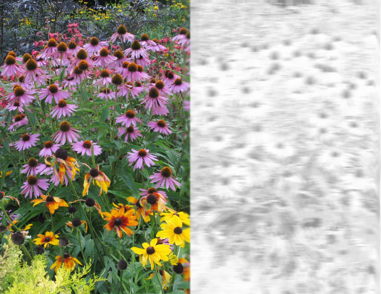
 Patients with achromatopsia do not see any colors, they have blurred vision and are very sensitive to glare. Photo and simulation: Stylianos Michalakis
What is the primary aim of the clinical study in which you are now involved?
Martin Biel: The goal is to effectively treat, i.e. remedy the principal symptoms associated with, a hitherto incurable visual disorder known as achromatopsia.
Stylianos Michalakis: Patients with complete achromatopsia, of which there are about 3000 in Germany, are daylight blind from birth. They are unable to distinguish colors, their visual acuity is severely impaired, and their eyes are hypersensitive to ambient light.
Biel: To cope with this photophobia, they have to wear very darkly toned sunglasses or contact lenses from an early age, as even very low levels of light cause them extreme discomfort. The disease is due to gene mutations, which result in the loss of an essential ion channel in the so-called cone cells that mediate color vision. We want to restore this function by introducing an intact copy of the gene into these photoreceptor cells.
What impact does this genetic error have?
Biel: The visual sense depends on the conversion of a physical stimulus, light, into a chemical reaction – hydrolysis of the molecule cyclic GMP (cGMP). The breakdown of cGMP is sensed by the ion channel and induces the channel to close, causing the membrane of the photoreceptor cell to become hyperpolarized. Thus light is transduced into a signal that triggers a cellular process. In patients with achromatopsia this ion channel is missing. They can perceive light because retain the photoreceptor protein, but they cannot process the signals in the normal way. So the information is not transmitted to the visual cortex of the brain, where the retinal image is normally reconstructed.
Project RD-Cure is designed by a consortium of clinicians and researchers based in Tübingen and Munich. What is your role in the collaboration?
Biel: We have worked closely with our colleagues in Tübingen for nearly 20 years now, not only on achromatopsia. We have studied the molecular operation of the ion channel in great detail. By carrying out targeted modification of the gene in mice, and demonstrating that these mice display the typical symptoms of color blindness, we have established an experimental model of achromatopsia. Together with our colleagues in Tübingen, we went on to develop a therapeutic strategy for the condition, and tested it in our model system. For this purpose, Dr. Michalakis constructed a set of viral vectors that were used to transport the intact gene into the mutant photoreceptors. These tests were so successful that we are now applying the method to patients. Here again, we have developed the gene vector and designed the viral vehicle to convey it into the target cells. The clinical part of the project – the introduction of the virus and the conduct of the clinical trial – lies in the hands of the team in Tübingen. The project is funded by the Tistou & Charlotte Kerstan Foundation.
How exactly is this type of gene therapy done?
Michalakis: Gene therapy basically involves the functional replacement of a defective gene. We will simply introduce the normal gene into the target cells. The endogenous gene is left untouched.
Biel: The defective gene stays where it is. Since it is inactive, it doesn’t affect the outcome for the patient. We just add an intact copy as a supplementary gene. It is not integrated into the genome and does not interfere with the action of the other genes. It remains outside the genome itself, but is accessible for the machinery that controls gene expression, and responds to the factors that direct it to produce the blueprint for synthesis of the missing ion channel.
Michalakis: In order to get the gene into the cone cells in the retina, we make use of an Adeno-Associated Virus (AAV) as a transport vehicle. We alter the viral genome by inserting the CNGA3 gene that codes for the ion channel, together with the regulatory sequences necessary for its expression in cone cells. We normally construct these “recombinant” viruses in the laboratory, but the virus used in the clinical study was produced by a subcontracted pharmaceutical company.
What makes this vector particularly suitable for transformation of the defective cone cells?
Michalakis: Our targets in the retina are sensory nerve cells. This already rules out a whole slew of gene transfer vectors, because they are unable to target neurons. AAV vectors, on the other hand, are ideal for this task, because they recognize and bind to specific surface receptors on nerve cells. They also express the genes they carry very efficiently and they do not get stuck in the extracellular matrix in the sub-retinal region, which surrounds the photoreceptors. – These features are all crucial for successful transduction of the intact gene.
How safe is the viral vector?
Biel: Adeno-Associated Viruses have been widely studied by many research groups worldwide. In fact, one form of AAV-based gene therapy has already been approved for clinical use. In the light of what we know, AAVs are exceptionally safe. They are not known to cause any form of disease in humans, and they do not integrate into the genome. So they cannot alter the structure of our genes – unlike retroviruses, which have been used for the construction of gene vectors in other contexts. So they cannot intervene in the normal business of the cell nucleus. They provide a supplementary function. They direct the synthesis of a product which the cell cannot otherwise fabricate, but they do not alter the cell’s biological program or character.
Michalakis: Furthermore, in our recombinant viruses virtually the whole viral genome has been replaced by the so-called expression cassette – the cargo or payload carried by the viral vehicle. Key elements required for viral replication have been deleted, so the viruses cannot reproduce in the body. A further safeguard lies in the fact that the viruses are introduced locally. They are introduced into the eye, which is a closed system. That reduces the risk that they will induce an immune response, which is something that must be reckoned with whenever viruses or foreign DNAs are systemically, rather than locally, introduced into the body. In our case, the viruses and the DNA they carry with them cannot escape from the site of application. They are restricted to a very small compartment below the retinal epithelium.
How is the treatment carried out in practice? What does it involve for the patient?
Biel: It’s basically a minimal surgery, which is carried out under anesthesia. A very fine capillary is used to inject a certain number of virus particles under the retina. At the site of injection, the retinal epithelium is slightly elevated and forms a tiny bleb. The viruses are taken up by the cells in the vicinity and after a time the bleb disappears. This latter process can be non-invasively monitored.
And a single injection is sufficient?
Michalakis: A single injection should be sufficient to ensure lifelong expression of the intact gene in the photoreceptor cells. The primary aim of the current Phase I study with 9 patients is to ensure that the approach is safe. The other main goal is to determine the most effective dosage level. In addition, we have defined a number of secondary endpoints, which will provide indications of the efficacy of the treatment.
What happens next?
Michalakis: For a so-called ATMP (Advanced Therapy Medicinal Product), to which gene gene therapies belong, the study design is relatively economical. We may be able to proceed directly to a Phase III study, but a phase I trial is usually followed by a phase II study, in which a larger number of patients is treated with what is deemed to be the optimal dosage.
Is this approach translatable in principle to other visual disorders?
Michalakis: About 200 genes have been identified which, when mutated, lead to monogenetic visual diseases, and doubtless many more remain to be discovered. Defects in any of 50 different genes can result in retinitis pigmentosa, a degenerative disease in which specific retinal photoreceptors die. Six genes have been linked to achromatopsia, and all of them should be amenable to the approach we are using to correct for defects in the CNGA3 gene.
Biel: But in some of these genes, different mutations give rise to different disease conditions, depending on their position within the gene. So the figure of 200 genes is likely to be a conservative estimate. In principle, introducing a copy of the intact gene into the cells directly affected should be applicable to every retinal disorder that is attributable to a defect in a single gene.
Michalakis: The consortium is now planning a second clinical study, which will get underway in about 18 months and will be devoted to retinitis pigmentosa (RP). In the case of this disease, for which there is no effective treatment so far, the cells that are primarily affected are not the cone cells but the rods. Because rod cells are required only for vision in dim light, – in other words, for night vision – many patients with RP initially notice little deterioration. However, over time, more and more rod cells degenerate and die. And at some point, the cone cells, although they are not directly affected by the RP mutation, begin to degenerate also, and daylight vision becomes progressively restricted. First peripheral vision is lost, and the visual field continues to shrink, which produces so-called tunnel vision. A significant fraction of these patients become clinically blind between the ages of 40 to 50. Here, the idea is to intervene early to save as many rod cells as possible and thus prevent secondary degeneration of the vital cones.
Prof. Dr. Martin Biel holds the Chair of Pharmacology for Natural Sciences at LMU. Dr. Stylianos Michalakis is Privatdozent and Junior Research Group Leader at the Chair of Pharmacology for Natural Sciences at LMU.