
STORM / PALM
Whereas the resolution of a conventional optical microscope is limited to ~200-300 nm due to diffraction of light on the aperture of the microscope, super-resolution methods like Stochastic Optical Reconstruction Microscopy (STORM) and Photoactivation Localization Microscopy (PALM) allow an increase of optical resolution by a factor of 10 up to ~20 nm in lateral and ~50 nm in axial direction.
Both methods rely on the usage of fluorescent emitters that can be stochastically switched between a bright and a dark state so that only one fluorescent emitter in a diffraction limited spot is active at a time. In this case, it is possible to calculate the positions of the distinct fluorophores with high accuracy, e.g. by fitting the resulting point spread function to a 2D Gaussian function. At the end , a high-resolution image can be rendered out of all the obtained localizations.
Our setup is able to perform both STORM and PALM, where the difference between methods lies in the nature of the fluorescent label, which are either photoswitchable organic dyes (e.g. Alexa Fluor 647, Cy5) for STORM or photoswitchable fluorescent proteins for PALM.
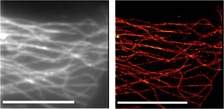
The following figure demonstrates the gain in resolution obtained by STORM at the example of HeLa cell microtubules stained with fluorescently labelled antibodies: the left panel shows the image obtained by conventional widefield microscopy in opposite to the right panel showing the corresponding high-resolution STORM image (bars = 10 µm):
Recommended literature:
- Rust M, et al. (2006) Sub-diffraction-limit imaging by stochastic optical reconstruction microscopy (STORM). Nature Methods 3(10): 793-795.
- Betzig E, et al. (2006) Imaging Intracellular Fluorescent Proteins at Nanometer Resolution. Science 313(5793): 1642-1645.
- Huang B, et al. (2008) Three-Dimensional Super-Resolution Imaging by Stochastic Optical Reconstruction Microscopy. Science 319(5864): 810-813.
- Heilemann M, et al. (2008) Subdiffraction-Resolution Fluorescence Imaging with Conventional Fluorescent Probes. Angew. Chem. Int. Ed. 47(33): 6172-6176.